May 4, 11:00am
Abstract:
Even though structured-light triangulation is a decades-old
problem, much remains to be discovered about it---with potential
ramifications for computational imaging more broadly.
I will focus on two specific aspects of the problem that are influenced by
recent developments in our field. First, programmable coded-exposure sensors
vastly expand the degrees of freedom of an imaging system, essentially
redefining what it means to capture images under structured light. I will
discuss our efforts to understand the theory and expanded capabilities of such
systems, and to build custom CMOS sensors that realize them. Second, I will
outline our recent work on turning structured-light triangulation into an
optimal encoding-decoding problem derived from first principles. This opens
the way for adaptive systems that can learn on their own how to optimally
control their light sources and sensors, and how to convert the images they
capture into accurate 3D geometry.
 Speaker bio:
Kyros Kutulakos is a Professor of Computer Science at the University of
Toronto. He received his PhD degree from the University of Wisconsin-Madison in
1994 and his BS degree from the University of Crete in 1988, both in Computer
Science. Kyros has been a pioneer in the area of computational light transport,
developing theoretical tools and computational cameras to analyze light
propagation in real-world environments. He is the recipient of an Alfred P.
Sloan Fellowship, an Ontario Premier's Research Excellence Award, a Marr Prize
in 1999, a Marr Prize Honorable Mention in 2005, and four other paper awards
(CVPR 1994, ECCV 2006, CVPR 2014, CVPR 2017). He was Program Co-Chair of CVPR
2003 and ICCV 2013, and also served as Program Co-Chair of the second ICCP
conference back in 2010.
Speaker bio:
Kyros Kutulakos is a Professor of Computer Science at the University of
Toronto. He received his PhD degree from the University of Wisconsin-Madison in
1994 and his BS degree from the University of Crete in 1988, both in Computer
Science. Kyros has been a pioneer in the area of computational light transport,
developing theoretical tools and computational cameras to analyze light
propagation in real-world environments. He is the recipient of an Alfred P.
Sloan Fellowship, an Ontario Premier's Research Excellence Award, a Marr Prize
in 1999, a Marr Prize Honorable Mention in 2005, and four other paper awards
(CVPR 1994, ECCV 2006, CVPR 2014, CVPR 2017). He was Program Co-Chair of CVPR
2003 and ICCV 2013, and also served as Program Co-Chair of the second ICCP
conference back in 2010.
May 5, 11:00am
Abstract:
Although treatment algorithms vary, surgery is the primary treatment modality for
most solid cancers. The fundamental goal of a curative oncologic surgery is complete
cancer removal but equally important from the patient’s point of view is function
preservation and pain control. Currently, there is an increasing trend to adopt
minimally invasive technologies such as endoscopic and robotic surgery in order to
decrease patient morbidity. However, the ability for surgeons to distinguish between
normal and diseased tissue by texture or consistency is significantly reduced with these
techniques (due to lack of ability to physically touch the tissue) and is limited to visual
inspection alone. Using white light reflectance which is the current standard mode of
illumination in operating rooms, the visual difference between normal and cancerous
tissue can be imperceptible. The inability of surgeons to visually distinguish between
tumor and normal tissue leads to residual cancer cells left behind at the edges of resection,
i.e. positive surgical margins and can be as high as 20-40% in breast cancer lumpectomy,
21% for radical prostatectomy, and 13% for HNSCC.
Molecular imaging with fluorescence provides enhanced visual definition between diseased and
normal tissue and have been shown to decrease PSM in both animal models and patients.
Molecular imaging with fluorescence can also provide enhanced visualization of important
structures such as nerves to improve preservation and minimize inadvertent injury.
Our laboratory has extensive experience in development of both nerve and tumor injectable
markers for surgical visualization. In presentation we will discuss the development of nerve
and tumor markers combinations to improve intraoperative visualization – aka color-coded surgery.
 Speaker bio:
Dr. Nguyen is a Professor in the Department of Surgery at the
University of California San Diego (UCSD). She received her combined MD/PhD degree
from Washington University, School of Medicine in St. Louis, MO. She completed her
General Surgery Internship at Barnes Jewish Hospital in St. Louis and residency in
Head and Neck Surgery and subspecialty fellowship training in Neurotology/Skull Base
Surgery at UCSD. She is board certified in both Head and Neck Surgery and
Neurotology/Skull Base Surgery and is the fellowship director for the ACGME
accredited fellowship in Neurotology/Skull Base Surgery at UCSD.
Speaker bio:
Dr. Nguyen is a Professor in the Department of Surgery at the
University of California San Diego (UCSD). She received her combined MD/PhD degree
from Washington University, School of Medicine in St. Louis, MO. She completed her
General Surgery Internship at Barnes Jewish Hospital in St. Louis and residency in
Head and Neck Surgery and subspecialty fellowship training in Neurotology/Skull Base
Surgery at UCSD. She is board certified in both Head and Neck Surgery and
Neurotology/Skull Base Surgery and is the fellowship director for the ACGME
accredited fellowship in Neurotology/Skull Base Surgery at UCSD.
Her clinical practice is at UCSD Health Systems where she cares for patients
with diseases of the facial nerve, ear and skull base. She has subspecialty interest
in facial nerve reanimation and supportive procedures for patients with facial paralysis
including nerve substitutions, cross facial, platinum eyelid weight placement, lower lid
ectropion repair, brow ptosis repair and temporalis tendon transfer. Additionally, she is
also able to provide non-surgical options including botox and fillers to support patients
with facial paralysis. She also specializes in hearing restoration surgeries including
cochlear implantation.
Dr. Nguyen’s interest in molecular imaging for surgical navigation began during her fellowship
at UCSD where she collaborated with Dr. Roger Tsien (1952-2016), Nobel Laureate, Chemistry 2008.
She has been awarded the Presidential Early Career Award for Scientists and Engineers (PECASE, April 2014).
The Presidential Award is the highest honor bestowed by the U.S. government on outstanding
scientists and engineers beginning their independent careers.
May 6, 11:00am
 Abstract:
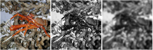
The appearance of visual scenes and the information that can be extracted from
them naturally depends on the characteristics of the sensor. Animal visual systems
vary significantly in a number of functional parameters including: spatial and temporal
resolution, spectral sensitivity and dimensionality of color space, and the ability to
discriminate both linear and circular polarization. Our position as humans is both privileged
and limited in that we have very high spatial resolution, but limited spectral range, relatively
poor color vision, and no useful polarization sensitivity. Therefore our view, and the
view of many of our imaging systems, is not typically representative of what other animals see.
Computational power and our knowledge of animal visual systems has now risen to the point
where we can model how the world looks to other species, which has opened doors to further
investigation in multiple fields, both basic and applied. This talk uses several examples
from the marine world to make the general point that how an animal (or a human) sees
the world profoundly affects how it can interact with it.
Abstract:
The appearance of visual scenes and the information that can be extracted from
them naturally depends on the characteristics of the sensor. Animal visual systems
vary significantly in a number of functional parameters including: spatial and temporal
resolution, spectral sensitivity and dimensionality of color space, and the ability to
discriminate both linear and circular polarization. Our position as humans is both privileged
and limited in that we have very high spatial resolution, but limited spectral range, relatively
poor color vision, and no useful polarization sensitivity. Therefore our view, and the
view of many of our imaging systems, is not typically representative of what other animals see.
Computational power and our knowledge of animal visual systems has now risen to the point
where we can model how the world looks to other species, which has opened doors to further
investigation in multiple fields, both basic and applied. This talk uses several examples
from the marine world to make the general point that how an animal (or a human) sees
the world profoundly affects how it can interact with it.
 Speaker bio:
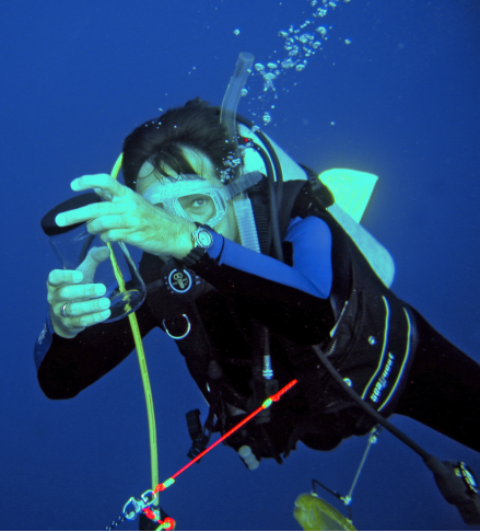
Originally trained in mathematics and art, Sönke Johnsen has studied camouflage, signaling,
and non-human visual modalities for the last 30 years. He is particularly interested in
vision and camouflage in the open ocean, but has also worked on coastal and terrestrial species,
magnetoreception, nocturnal illumination, and human cataracts. His research combines mathematical
analyses with behavioral and morphological studies and in situ measurements and observations.
His field work primarily involves open-ocean research cruises that use SCUBA and deep-sea submersibles.
In addition to exploring the evolution and diversity of the optical and visual tricks that animals perform,
Professor Johnsen is interested in improving communication between theoretical and experimental scientists,
biologists and physicists, and scientists and artists. Outreach is a strong focus, and Johnsen’s research
has been presented in numerous magazines, newspapers and television shows. Professor Johnsen has also
written two books, The Optics of Life and Visual Ecology, and is currently working on a third for
a lay audience. In his spare time, he is an avid nature photographer and amateur farmer.
Speaker bio:
Originally trained in mathematics and art, Sönke Johnsen has studied camouflage, signaling,
and non-human visual modalities for the last 30 years. He is particularly interested in
vision and camouflage in the open ocean, but has also worked on coastal and terrestrial species,
magnetoreception, nocturnal illumination, and human cataracts. His research combines mathematical
analyses with behavioral and morphological studies and in situ measurements and observations.
His field work primarily involves open-ocean research cruises that use SCUBA and deep-sea submersibles.
In addition to exploring the evolution and diversity of the optical and visual tricks that animals perform,
Professor Johnsen is interested in improving communication between theoretical and experimental scientists,
biologists and physicists, and scientists and artists. Outreach is a strong focus, and Johnsen’s research
has been presented in numerous magazines, newspapers and television shows. Professor Johnsen has also
written two books, The Optics of Life and Visual Ecology, and is currently working on a third for
a lay audience. In his spare time, he is an avid nature photographer and amateur farmer.